Introduction
Vascular surgery today, while advanced, relies on separate tools for visualizing anatomy and measuring blood flow, leaving room for uncertainty. Imagine an instrument that integrates Transit Time Flow Measurement (TTFM) with high-frequency imaging, transforming vascular surgeries with precision and real-time insights, guiding you performing surgery with best possible patient outcomes.
The MiraQ is the only device combining TTFM and high-resolution imaging in one unit. It empowers vascular surgeons with precision and real-time insights, elevating patient outcomes in one innovative solution. Perform vascular imaging, measure blood flow and receive immediate feedback on how well a graft is functioning and minimize the risk for reinterventions.
With graft patency being the predominant predictor of long-term survival after vascular surgery, surgeons can ensure improved patient outcomes.
Peripheral Bypass Surgery
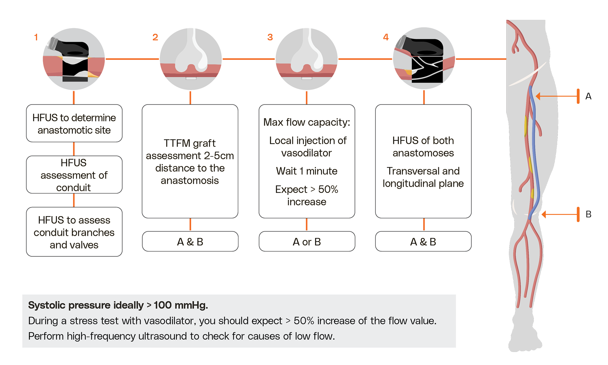
Volumetric blood flow values have proven to be an important factor for predicting graft longevity. Good graft function increases the likelihood of saving the patient’s leg. The risk of early postoperative occlusion is significantly increased if the intraoperative flow volume after open bypass revascularization is lower than comparable flow reference values or an additional stress test performed with a vasodilator is not showing expected increase in flow.
Assessment of anastomotic sites using Medistim's imaging probe which is especially adapted for intraoperative use, can reveal otherwise unseen imperfections inside the vessels. The Medistim technology gives surgeons the chance to correct technical errors or defects before closure.
With graft patency being the predominant predictor of long-term survival after vascular surgery, surgeons can ensure improved patient outcomes using this unique quality assessment device.
At sites A+D:
Perform imaging to determine the anastomotic sites.
At sites B+C:
Measure flow volume in the graft.
At sites B+C:
Perform stress test with vasodilator and expect >50% flow increase.
At sites A+D:
Perform imaging of the anastomoses to reveal any restrictions.
Carotid Endarterectomy (CEA)
The CIDAC Study
Intraoperative completion imaging after CEA has the benefit of reducing the risk of perioperative stroke. The CIDAC study concludes that intraoperative duplex ultrasound (IDUS) enables more correct detection of higher-grade defects than angiography (p<0.001). Defects considered for revision were detected in 22 vs. 10 patients for IDUS vs. angio (p=0.04). The inter-observer reliability of IDUS is higher than of angio (p=0.003).
2023 ESVS Guidelines
A section of the ESVS Guidelines describes monitoring and quality control after CEA to identify luminal thrombus after flow restoration, diagnose intimal flaps and diagnose residual stenoses during surgery. The recommendation is based on a meta-analysis by Knappich et al. from 2021 showing that both ultrasound imaging and angiography are associated with a reduced risk of death and stroke after CEA. The CIDAC study contributes to the conclusion of this meta-analysis.
Recommendation 77
For patients undergoing carotid endarterectomy, intra-operative completion imaging with angiography, duplex ultrasound or angioscopy should be considered in order to reduce the risk of peri-operative stroke.
Class IIa, Level B
References: Knappich et al. (2021)
At sites A+D:
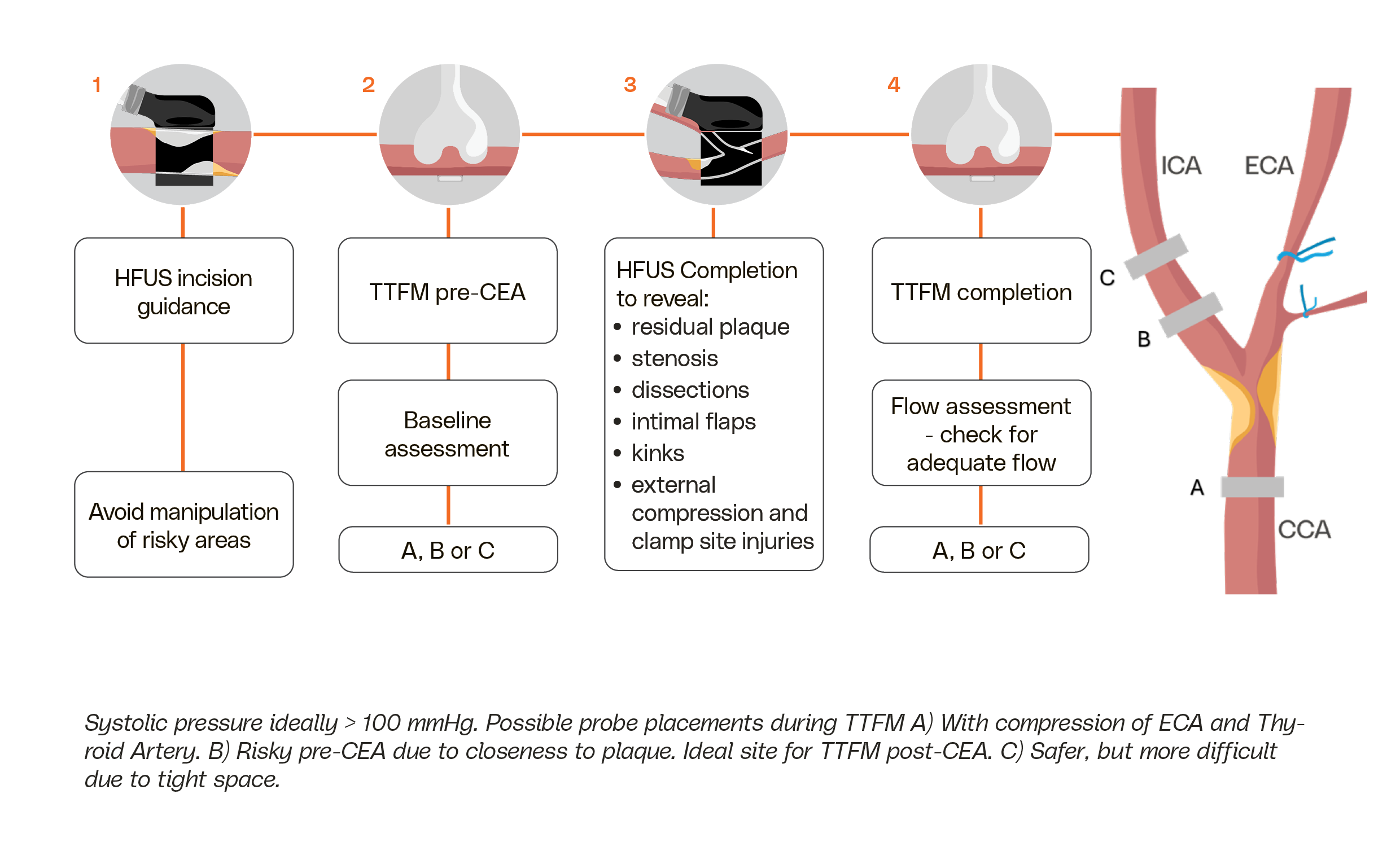
Perform intraoperative imaging to guide incision and assess the size of the plaque.
At sites B+C:
Optional:
At sites A, B or C:
Assess flow to establish baseline.
At sites B+C:
Perform imaging completion control:
- Check for residual intimal flaps or plaque.
- Check clamping sites for injury.
At sites A+D:
Optional:
TTFM completion control at sites B or C:
- Assess flow to avoid cerebral hyperperfusion syndrome.
- Check for adequate flow especially through kinked arteries.
Ultrasound reduces the risk of in-hospital death and stroke by 26%, showing higher sensitivity compared to angiography.
Dr. Christoph Knappich, Klinikum rechts der Isar der Technischen Universität München (TUM)
AV Access Surgery
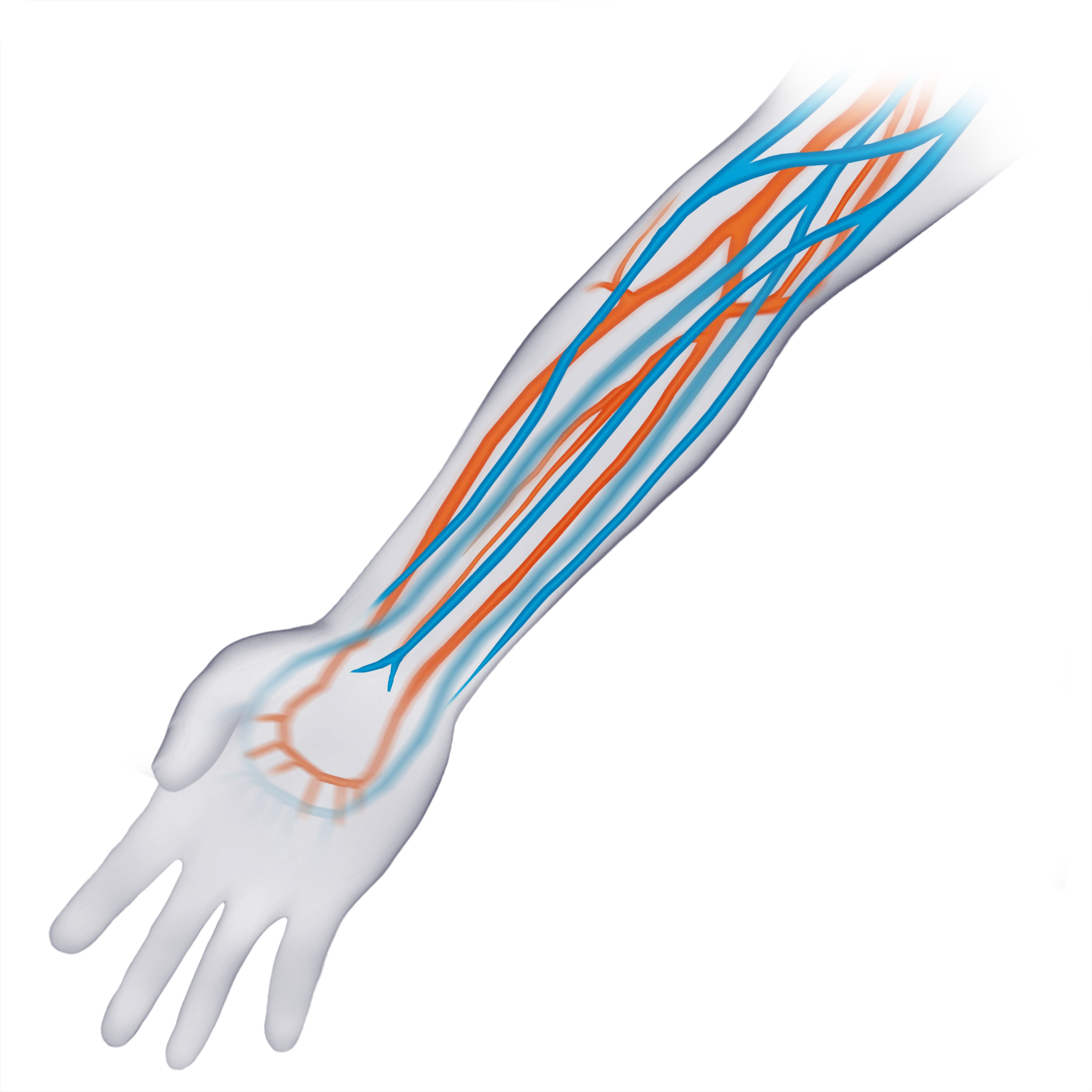
If the flow in the fistula is too high, there is a risk of hand ischemia and heart failure. For patients experiencing high-flow issues, flow reduction using intraoperative flow monitoring is an effective and durable technique allowing for the correction of hand ischemia and cardiac insufficiency.
The MiraQ device enables successful fistula maturation contributing to a long lasting shunt and effective high-flow modulation improving the patient's quality of life.
At sites A+D:
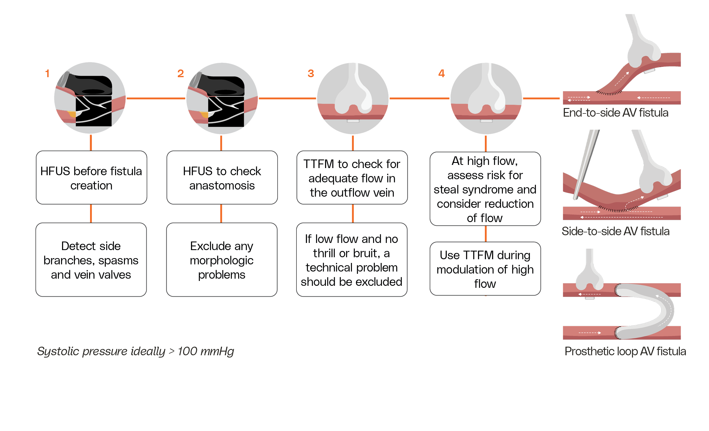
Perform imaging before fistula creation to detect significant side branches and valves.
At sites B+C:
Measure flow in the outflow vein to assess adequate flow.
At sites B+C:
Perform imaging to check the quality of the anastomosis and exclude any morphological issues.
At sites A+D:
Perform flow modulation with flow and imaging in high-flow cases during initial surgery or after maturation.